Since much of this thesis makes references to brain anatomy and function, this section deals with the terminology used in the discipline of neurology. There are four sections covering brain anatomy, brain cellular structure and brain function. Further reference can be obtained from the many good books on brain biochemistry and neuroanatomy [13,14,15].
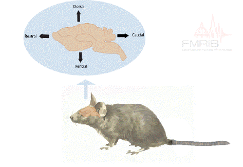
In order to describe the position of structures relative to each other in the brain, neurology has a number of terms of direction, many of which are derived from the Latin or Greek. There are two axes which describe the organisation of the central nervous system (CNS). These are most easily understood in animals with the spinal cord running horizontally rather than vertically. In this case the rostral-caudal axis runs from nose to tail, and the dorsal-ventral axis runs perpendicularly to this as shown in Figure 3.4a. Now using this system in the human spinal cord, 'top to bottom' is 'caudal to rostral', and 'back to front' is 'dorsal to ventral' (Figure 3.4b). In the human brain however these axes turn through 90 degrees, so the front of the brain is rostral, top is dorsal, and base is ventral.
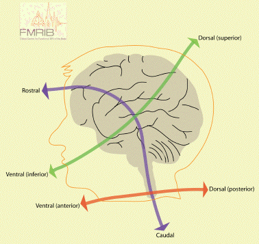
In addition to these labels, there is another perpendicular set of axes which is the same for spinal cord and brain, that is anterior=front, posterior=back, superior=top, inferior=bottom.
The midline runs down the centre of the brain, separating left from right. If two structures are on the same side of the midline, they are said to be ipsilateral, whereas they are contralateral if they are on opposite sides. When comparing two structures, the one closest to the midline is medial, as opposed to the other which is lateral.
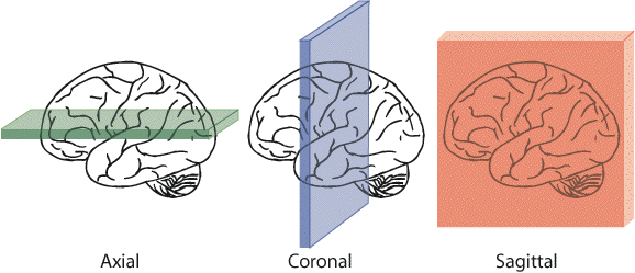
When viewing sections through the brain, three mutually perpendicular planes are commonly considered, as shown in Figure 3.5. These are axial (or transverse) coronal, and sagittal.
The neuron is the basic functional unit of the nervous system. The brain consists of several hundred billion neurons, communicating by billions of interconnections. All neurons consist of four distinct parts: cell body, dendrites, axons and axon terminals (Figure 3.6).
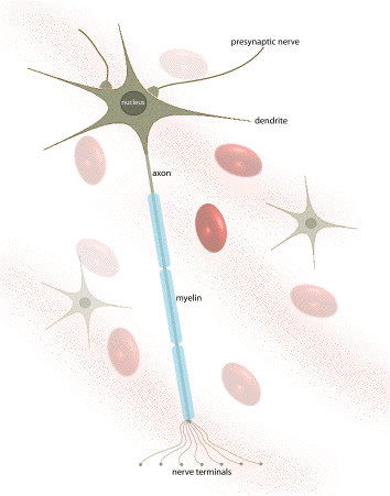
The cell body (or soma) contains the nucleus of the cell, as well as the essential cellular organelles, such as the energy generating mitochondria. The cell body has many branches, called dendrites, which receive signals from other cells, and are often covered in dendritic spines. Extending from the cell body in one direction is an axon. The length of axons can be several centimetres or longer. Axons carry information from one neuron to another, and are terminated at the synaptic knob, which is attached to the dendrites or cell body of another neuron. Signals are transferred across the synapse by means of a chemical neurotransmitter.
Signals travel along the axon by generating and propagating an action potential. This is produced by letting the delicate balance of sodium, potassium and chloride ions across the cell membrane be altered, thus generating an electrical signal that flows along the axon. If the axon is coated in a fatty sheath, called myelin, the signal travels at higher speeds of anything up to 120 ms-1. When the signal reaches the synapse, the synaptic knob emits a neurotransmitter which acts either to encourage the next neuron to 'fire' (excitatory) or discourage the neuron to fire (inhibitory).
The CNS contains a number of different types of neurons, which are tailored to the job they perform. Signals from sensory receptors over the body feed along the spinal cord to the brain, and signals are sent from the brain to make muscles contract. Many medical conditions are caused by the failure of the CNS to function correctly, for example in Parkinson's disease there is a deficiency of the neurotransmitter dopamine.
For every neuron in the CNS there are also ten glial cells. These cells provide support for neurons, for example the microglia which perform a scavenger role, and oligodendrocytes which form the myelin sheath around the axons.
The central nervous system consists of the spinal cord and the brain. The brain is then further divided into the forebrain, midbrain, and hindbrain (Figure 3.7).
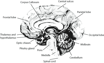
The largest region is the forebrain, which contains the cerebral hemispheres, the corpus callosum, thalamus, hypothalamus, and hippocampus. the hindbrain consists of the cerebellum, pons, and medulla. The structure of each of these is described below.
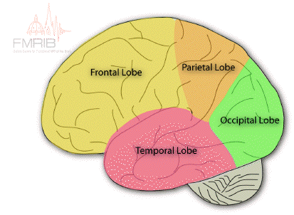
The cerebrum is divided into two hemispheres, the left and the right, separated by the longitudinal fissure. Anatomically these are identical in form, each being split into four lobes; the frontal lobe, the parietal lobe on the top, the temporal lobe on the side, and the occipital lobe at the back (Figure 3.8). The frontal and parietal lobes are separated by the central sulcus, and the temporal lobe separated by the lateral fissure. The corpus callosum joins left and right hemispheres.
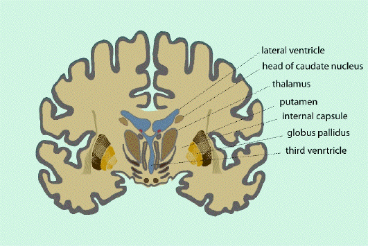
Under the surface of the cerebral hemispheres are bundles of fibres, the basal ganglia, connecting together many regions of the cerebral cortex. The thalamus, hypothalamus and hippocampus are located at the centre of the forebrain, just above the midbrain (Figure 3.10). At the rear of the brain is a more tightly folded structure called the cerebellum, which is connected to the pons, the medulla, and finally the spinal cord.
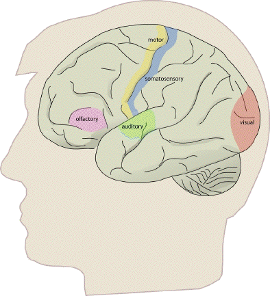
The functional organisation of much of the brain is poorly understood. However many of the regions involved in sensory and motor function have been identified.
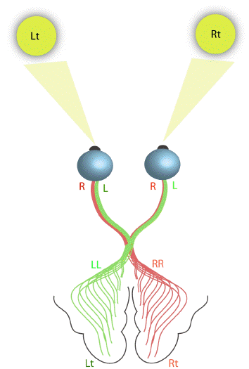
The primary visual cortex is located in the occipital lobe, which deals with the reception and interpretation of vision. The right visual field is mapped on to the left cerebral hemisphere, and the left visual field on to the right hemisphere (Figure 3.11). The signals from the retina travel along the optic tracts, which cross over at the optic chiasm.
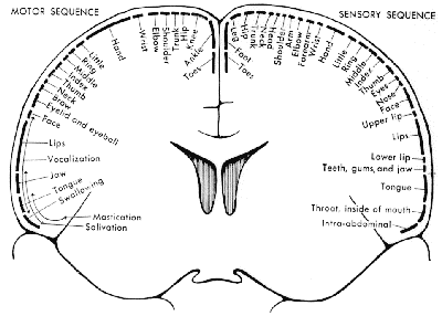
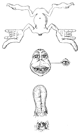
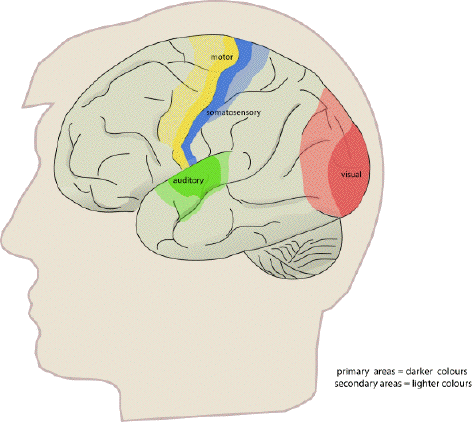
The organisation of the somatosensory cortex shows similarity to a map of the surface of the body[16]. This is illustrated in what is known as the sensory homunculus (Figure 3.13). A much greater part of the somatosensory cortex is associated with the hand and face, compared to regions not so important in tactile tasks such as the leg. Similarly there is a motor homunclus which illustrates the layout of the motor cortex. The hand is given much more cortical surface in the motor cortex than in the somatosensory cortex, representing the highly sophisticated tasks that the hands perform.
All the regions mentioned so far are primary cortices because they are most closely involved with the brain's input and output. Much of the rest of the brain is given over to integrating these stimuli, and interpreting how to respond. The regions responsible for these more abstract tasks are termed secondary (Figure 3.14). For example, the secondary motor regions, which are more anterior in the frontal lobes to the primary motor cortex, are responsible for planning and initiating motion, and the secondary visual area, close to the primary visual cortex is involved in interpreting colours and movement in the visual information. The tertiary areas, or association cortices, are responsible for the higher brain functions such as interpretation and memory.
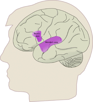
Some specific regions of interest are those responsible for speech, which are located in the left hemisphere of most people. Broca's area is in the lower part of the frontal lobe (Figure 3.15), and is involved in the formation of sentences, and Wernicke's area, located in the temporal lobe is involved in the comprehension of speech.
This is just a brief sketch of brain function as it is understood at present. New literature is appearing at a rapid pace, confirming and evolving the models. The motor system is covered in more detail in Chapter 7.