


Next: Estimation
Up: Local Parameter Estimation
Previous: Increasing the Complexity -
Local Parameter Estimation: Methods
Data acquisition DT-MRI datasets were acquired on a single
healthy volunteer. The images were obtained on a 3.0 T Varian Inova
scanner using a diffusion-weighted single-shot EPI sequence. To
minimize eddy currents, a doubly-refocused spin-echo sequence was
implemented [17]. A birdcage radio-frequency head coil
was used for both pulse transmission and signal detection. The
diffusion gradients achieved a maximum gradient strength of 22
 . Each data set consisted of 3 non-diffusion-weighted and
60 diffusion-weighted images acquired with a b-value of 1000
. Each data set consisted of 3 non-diffusion-weighted and
60 diffusion-weighted images acquired with a b-value of 1000
 . The diffusion gradients were uniformly distributed through
space using the optimized scheme proposed by Jones [18].
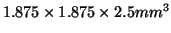
Each set of images contained 42 contiguous slices with a 2.5
. The diffusion gradients were uniformly distributed through
space using the optimized scheme proposed by Jones [18].
Each set of images contained 42 contiguous slices with a 2.5  thickness. A half k-space acquisition was performed with a matrix size
set to
thickness. A half k-space acquisition was performed with a matrix size
set to
 and a field of view of
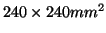 . The
images were interpolated to achieve a matrix size of
. The
images were interpolated to achieve a matrix size of
 and a final resolution of
 . To
minimize motion artifacts, peripheral gating was used such that
triggering occurred on every cardiac cycle. The echo time was set to
106 ms while the effective repetition time was 14 R-R intervals. The
total scan time for each dataset was approximately 15 minutes,
depending on heart rate.
. To
minimize motion artifacts, peripheral gating was used such that
triggering occurred on every cardiac cycle. The echo time was set to
106 ms while the effective repetition time was 14 R-R intervals. The
total scan time for each dataset was approximately 15 minutes,
depending on heart rate.
Subsections
Next: Estimation
Up: Local Parameter Estimation
Previous: Increasing the Complexity -
Tim Behrens
2004-01-22