


Next: Results
Up: Motion Correction
Previous: Motion Correction
As the intensity values are of great interest after motion correction,
attention must be paid to not only the estimation but the application
of the transformation. Interpolation probably has the largest impact
on the quality of the transformed data, with sinc interpolation
methods often being used, although no absolute consensus on the best
method exists. However, the loss of information outside the FOV,
usually seen in the end slices, can also be very detrimental to the
final statistical maps in these areas.
Our motion correction implementation has also been designed to handle the potentially problematic
issue of end-slice interpolation. It is frequently the case that under even small affine motion of the head, voxels in
the top and bottom slices can move either in or out of the field
of view (see Figure 7). Other schemes approach this
by either assuming that all affected voxels are zero (AIR) or can be
completely excluded from further calculations (SPM). This clearly impacts later analysis as valuable spatial information may
be lost.
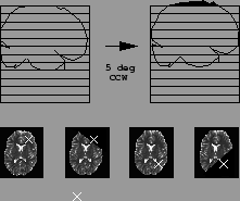
Figure 7:
Top row: demonstration of a significant section of brain moving out of the FOV (depicted as a stacked-slice grid) when the patient exhibits 5 pitch about the center of the brain. Bottom row: Corresponding voxels in adjacent scans (shown as white crosses) can be lost if there is appreciable movement of brain in the end slices. Diagram shows reference brain and corrected slice which has moved out of the FOV because of patient motion for two movements
 |
We counter this situation by padding the end-slices when applying the
estimated transformation (i.e. increasing the extent of
each volume by 2 slices). This means that if data is to be
interpolated from outside the FOV, it will take on 'sensible' values (Personal Communication with Roger Woods, 1999).
Next: Results
Up: Motion Correction
Previous: Motion Correction
Peter Bannister
2002-05-03